Art with DNA – Digitally creating 16 million colors by chemistry
17. October 2023Previous limitation to 256 colors far exceeded
The DNA double helix is composed of two DNA molecules whose sequences are complementary to each other. The stability of the duplex can be fine-tuned in the lab by controlling the amount and location of imperfect complementary sequences. Fluorescent markers bound to one of the matching DNA strands make the duplex visible, and fluorescence intensity increases with increasing duplex stability. Now, researchers at the University of Vienna succeeded in creating fluorescent duplexes that can generate any of 16 million colors – a work that surpasses the previous 256 colors limitation. This very large palette can be used to "paint" with DNA and to accurately reproduce any digital image on a miniature 2D surface with 24-bit color depth. This research was published in the Journal of the American Chemical Society.
The unique ability of complementary DNA sequences to recognize and assemble as duplexes is the biochemical mechanism for how genes are read and copied. The rules of duplex formation (also called hybridization) are simple and invariable, making them predictable and programmable too. Programming DNA hybridization allows for synthetic genes to be assembled and large-scale nanostructures to be built. This process always relies on perfect sequence complementarity. Programming instability vastly expands our ability to manipulate molecular structure and has applications in the field of DNA and RNA therapeutics. In this novel study, researchers at the Institute of Inorganic Chemistry at the University of Vienna showed that controlled hybridization can result in the creation of 16 million colors and can accurately reproduce any digital image in DNA format.
A canvas the size of a fingernail
To create color, different small DNA strands linked to fluorescent molecules (markers) that can emit either red, green or blue color are hybridized to a long complementary DNA strand on the surface. To vary the intensity of each color, the stability of the duplex is lowered by carefully removing bases of the DNA strand at pre-defined positions along the sequence. With lower stability comes a darker shade of color, and fine-tuning this stability results in the creation of 256 shades for all color channels. All shades can be mixed and matched within a single DNA duplex, thus generating 16 million combinations and matching the color complexity of modern digital images. To achieve this level of precision in DNA-to-color conversion, > 45,000 unique DNA sequences had to be synthesized.
To do so, the research team used a method for parallel DNA synthesis called maskless array synthesis (MAS). With MAS, hundreds of thousands of unique DNA sequences can be synthesized at the same time and on the same surface, a miniature rectangle the size of a fingernail. Since the approach allows the experimenter to control the location of any DNA sequence on that surface, the corresponding color can also be selectively assigned to a chosen location. By automating the process using dedicated computer scripts, the authors were able to transform any digital image into a DNA photocopy with accurate color rendition. "Essentially, our synthesis surface becomes a canvas for painting with DNA molecules at the micrometer scale", says Jory Lietard, PI in the Institute of Inorganic Chemistry.
Resolution is currently limited to XGA, but the reproduction process is applicable to 1080p, as well as potentially 4K image resolution. "Beyond imaging, a DNA color code could have very useful applications in data storage on DNA", says Tadija Kekić, PhD candidate in the group of Jory Lietard. As evidenced by the 2023 Nobel Prize attributed to the development of quantum dots, the chemistry of color has a bright future ahead.
This work was financially supported by the Austrian Science Fund (FWF projects I4923, P34284, P36203 and TAI687).
Original publication:
T. Kekić, J. Lietard: A Canvas of Spatially Arranged DNA Strands that Can Produce 24-bit Color Depth. Journal of the American Chemical Society. Published online October 4, 2023.
DOI: 10.1021/jacs.3c06500
Pictures:
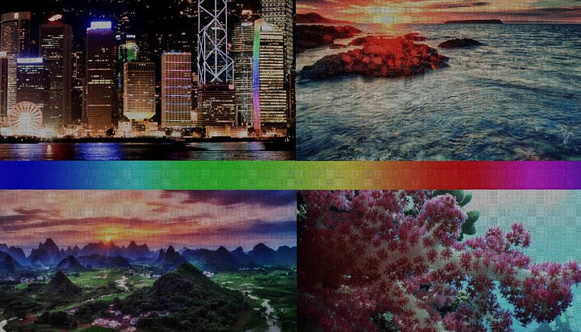
Fig. 1: The original digital image (in standard 24-bit color depth). C: cblee, Trey Ratcliff, stewartbaird and NOAA Ocean Exploration & Research
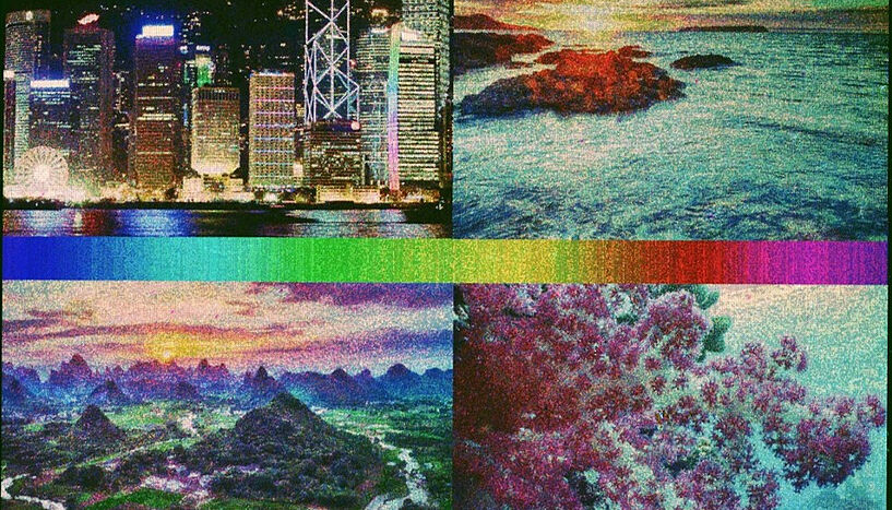
Fig. 2: The picture "photocopied" in DNA format by the authors. C: Tadija Kekic and Jory Lietard
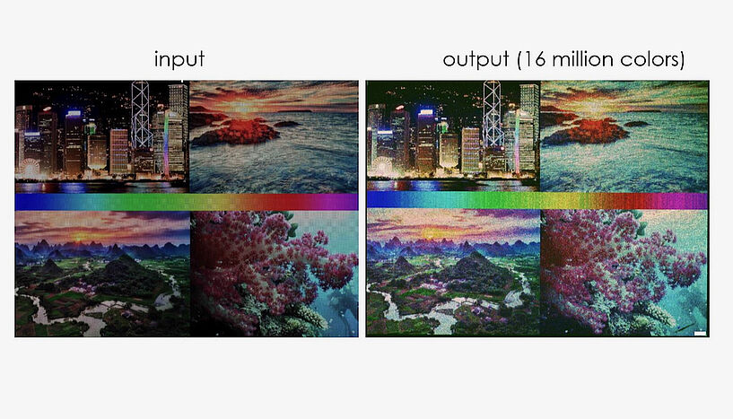
Fig. 3: Left: C: cblee, Trey Ratcliff, stewartbaird and NOAA Ocean Exploration & Research Right: C: Tadija Kekic and Jory Lietard
Scientific contact
Dr. Jory Lietard, PhD
Institut für Anorganische Chemie, Fakultät für ChemieUniversität Wien
1090 - Wien, Josef-Holaubek-Platz 2 (UZA II)
+43 1 4277 52646
jory.lietard@univie.ac.at
Further inquiry
Theresa Bittermann
Media Relations, Universität Wien1010 - Wien, Universitätsring 1
+43-1-4277-17541
theresa.bittermann@univie.ac.at
Downloads:
20231017_Lietard_Abb1_01.jpg
File size: 252,46 KB
20231017_Lietard_Abb2_01.jpg
File size: 308,69 KB
20231017_Lietard_Abb3_01.JPG
File size: 360,38 KB


